Real-time polymerase chain reaction

qPCR — CORE-ABR molecular panel

Laboratory-developed method

Developed and validated in house by Clinicore. Reviewed and approved by the Medical Director before use in patient testing.

Results from a laboratory-developed method are method-specific and are not directly comparable with another laboratory’s. That is the point of it — where a cleared assay answers the question adequately, Clinicore uses one.

How it works

Organisms are detected and quantified; resistance genes are reported qualitatively as detected or not detected. Reports are generated by CORE-ABR, which applies organism-aware antibiotic ranking incorporating intrinsic biology, detected resistance genes, and site-specific rules.

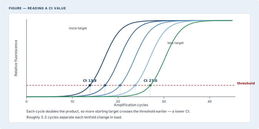
Five amplification curves from samples containing progressively less target DNA. Fluorescence rises as each cycle doubles the amplified product. The cycle at which a curve crosses the detection threshold is its Ct value. A sample containing more target crosses earlier and has a lower Ct; roughly 3.3 cycles separate each tenfold change in starting material.
Reading a Ct value.
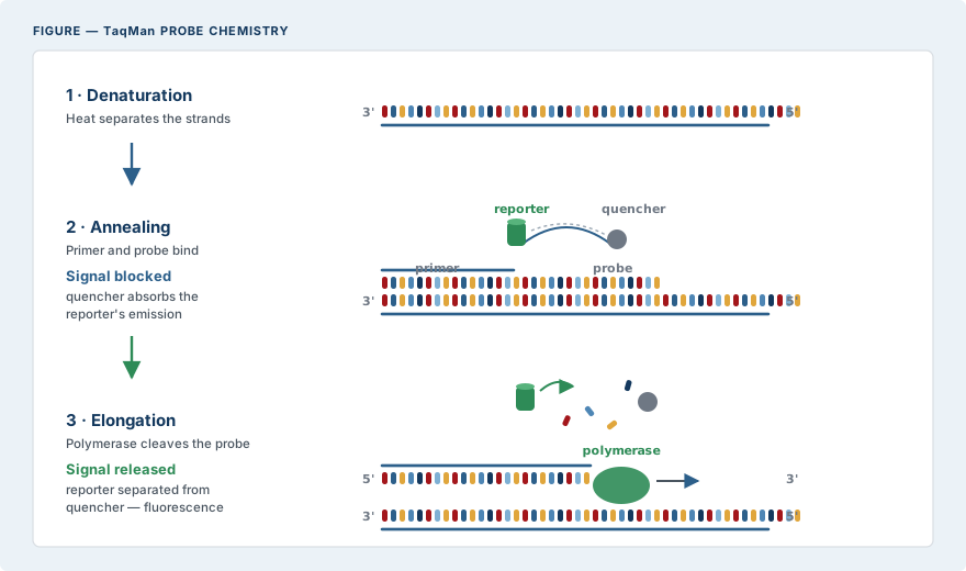
In denaturation, heat separates the two strands of the target DNA. In annealing, a primer binds the template and a sequence-specific probe hybridises downstream; the probe carries a reporter dye at one end and a quencher at the other, and while they remain close the quencher absorbs the reporter's emission so no signal is produced. In elongation, polymerase extends from the primer and cleaves the probe as it advances, separating reporter from quencher and releasing fluorescence. One probe cleaved equals one target copy amplified.
TaqMan probe chemistry — why the signal is specific to the intended sequence.
Panels on this platform