Clinical Science Review · Vol. 1 · For healthcare professionals
Unconjugated DHEA and DHEA-Sulfate Are Not Interchangeable
Abstract
- Rationale
- Across clinical practice, "DHEA status" is represented almost universally by dehydroepiandrosterone sulfate (DHEA-S), an analyte selected for its analytical convenience. DHEA-S, however, is a biologically inert sulfoconjugate, whereas unconjugated dehydroepiandrosterone (DHEA) is the receptor-active, intracrine and neuroactive species.
- Key distinctions
- The two pools differ by roughly two to three orders of magnitude in concentration and diverge sharply in half-life and temporal behaviour: DHEA-S is static, while DHEA is low, short-lived, and secreted in ACTH-driven pulses with diurnal structure. Because sulfotransferase (SULT2A1) and steroid sulfatase (STS) independently govern interconversion, circulating DHEA and DHEA-S can dissociate.
- Clinical implications
- Measurement of unconjugated DHEA enables assessments the sulfate cannot support — the cortisol:DHEA ratio as an index of hypothalamic–pituitary–adrenal balance, monitoring of DHEA replacement, and complete adrenal-androgen characterisation when paired with DHEA-S.
- Conclusion
- DHEA-S remains an appropriate screen for cumulative adrenal androgen output but is an incomplete surrogate for the active hormone. Paired measurement yields information neither analyte provides alone.
Keywords:dehydroepiandrosterone · DHEA-sulfate · adrenal androgens · SULT2A1 · steroid sulfatase · cortisol:DHEA ratio · intracrinology · LC-MS/MS
1. Background
DHEA and DHEA-S are the most abundant circulating steroids in humans, and both decline predictably across the adult lifespan — a decline reproducible enough that DHEA-S is treated as an endocrine biomarker of aging.1
In routine practice, an order for “DHEA” is fulfilled almost invariably by DHEA-S, because the sulfate circulates at high concentration and is stable throughout the day. That convention is analytically pragmatic, but it has quietly conflated measurability with completeness. The sulfate is easy to measure; it is not, on its own, a faithful readout of the active hormone.
This review sets out the biochemical, kinetic and clinical reasons to treat DHEA and DHEA-S as distinct analytes — and to measure the active hormone when the clinical question calls for it.
2. Two pools of one precursor
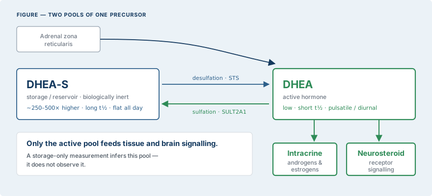
DHEA is synthesised in the adrenal zona reticularis and is reversibly sulfated to DHEA-S by cytosolic sulfotransferase SULT2A1; the reverse reaction, desulfation, is catalysed peripherally by steroid sulfatase (STS). DHEA-S is a hydrophilic circulating reservoir that is biologically inert until desulfation regenerates DHEA.
Only the unconjugated hormone is taken up and converted intracellularly — the intracrine pathway — into potent androgens and estrogens within target tissues, and only unconjugated DHEA acts as a neurosteroid at neuronal receptor sites.2
The clinical corollary is direct: the storage pool reports how much substrate exists; the active pool reports what is physiologically available now.
3. Static reservoir versus dynamic signal
The two analytes carry different temporal information.
DHEA-S has high concentration, a long half-life, and minimal diurnal variation — properties that make it a slow-moving, cumulative index.
Unconjugated DHEA behaves oppositely: present at far lower concentration, short half-life, secreted in pulsatile ACTH-driven bursts with clear diurnal and sex-specific structure.
In healthy older adults, nocturnal DHEA secretion is pulsatile and sexually dimorphic — and, critically, testosterone administration measurably lowered nocturnal DHEA while morning DHEA-S was unchanged.3
A static analyte cannot register a dynamic perturbation. The sulfate averages away exactly the signal the active hormone preserves.
4. Dissociation of the two analytes
Because SULT2A1 and STS are regulated independently — by tissue, age, disease state and pharmacologic exposure — the ratio of DHEA to DHEA-S is not fixed, and the two can move independently.
This is demonstrable empirically: in women with childhood sexual-abuse–related post-traumatic stress disorder, 24-hour DHEA-S differed from comparison groups while unconjugated DHEA did not.4
Where the analytes dissociate, a sulfate-only measurement discards the interconversion signal and can misrepresent active-hormone status. When two markers are capable of diverging, measuring one is a defined blind spot.
5. Applications favouring measurement of unconjugated DHEA
5.1 Cortisol:DHEA ratio and HPA-axis balance
The cortisol:DHEA ratio is used as an index of anabolic–catabolic balance and HPA-axis reactivity; because it requires the active hormone, it cannot be computed from the sulfate. Salivary DHEA and the cortisol:DHEA ratio have been applied to characterise acute stress responsiveness.5 Panels reporting only DHEA-S cannot furnish this index.
A note on units. Clinicore computes and reports the cortisol:DHEA ratio in DHEA units (ng/dL), with cortisol converted from µg/dL. Any published reference to this ratio must state the units it was computed on — laboratories reporting it in cortisol units produce values a thousandfold different, and the two are not comparable.
5.2 Monitoring DHEA replacement
Where DHEA is being replaced or supplemented, the unconjugated hormone is the physiologically relevant species to follow. DHEA replacement improves defined outcomes in adrenal insufficiency, with benefit most evident in women,6 and is an established consideration in the management of adrenal insufficiency.7
Notably, benefit is condition-specific: controlled data show no clear advantage of DHEA administration in eugonadal older men with a physiological age-related decline,8 underscoring that the value lies in measuring and interpreting, not in reflexive supplementation.
5.3 Complete adrenal-androgen characterisation
DHEA-S remains the appropriate marker of adrenal-derived androgen excess — roughly 20–30% of PCOS patients demonstrate adrenal androgen excess indexed by DHEA-S9 — and it assists in localising the source of androgen or cortisol excess in the evaluation of Cushing syndrome and adrenal tumours.10
Pairing DHEA-S with unconjugated DHEA adds the active-pool and interconversion dimension to that workup rather than replacing it.
5.4 Prognostic context
Low DHEA-S independently predicts all-cause and cardiovascular mortality in men in population cohorts.11
That prognostic evidence is specific to the sulfate. Unconjugated DHEA should be positioned as complementary physiological information, not as a validated substitute prognostic marker. Accurate framing preserves credibility.
6. Analytical considerations
The dominance of DHEA-S in laboratory ordering is, at root, a measurement-driven artifact. Its high concentration and stability make immunoassay straightforward and forgiving of pre-analytical variation.
Unconjugated DHEA is analytically more demanding: it circulates at substantially lower concentration, varies through the day, and is susceptible to immunoassay cross-reactivity — so reliable quantification favours a validated liquid chromatography–tandem mass spectrometry method with appropriate sensitivity and controlled specimen timing.
Laboratories able to quantify unconjugated DHEA to this standard therefore report information most panels omit by default.
Table 1. DHEA versus DHEA-S at a glance
| Property | DHEA (unconjugated) | DHEA-S (sulfate) |
|---|---|---|
| Biological role | Active precursor; intracrine and neurosteroid | Inert reservoir; requires desulfation |
| Relative concentration | Low | ~250–500× higher |
| Half-life | Short (hours) | Long (stable across the day) |
| Diurnal behaviour | Pulsatile, ACTH-driven, diurnal | Essentially flat |
| Information carried | Real-time adrenal output | Cumulative average output |
| Preferred assay | Validated LC-MS/MS | Immunoassay adequate |
| Distinct utility | Cortisol:DHEA ratio; therapy monitoring; active-pool status | Screening; aging index; prognostic cohorts |
7. Conclusions and recommendations
DHEA-S should not be treated as a proxy for the active hormone. A defensible ordering logic follows from the physiology:
- Screen with DHEA-S for cumulative adrenal androgen output.
- Add unconjugated DHEA when active-pool status, the cortisol:DHEA ratio, therapy monitoring, or analyte interconversion is clinically relevant.
- Interpret the two together. The relationship between the reservoir and the active hormone is itself informative, and it is invisible when only one is measured.
References
- Lane MA, et al. Dehydroepiandrosterone sulfate: a biomarker of primate aging slowed by calorie restriction. J Clin Endocrinol Metab. 1997;82(7):2093–96.doi:10.1210/jcem.82.7.4038
- Campbell B. Adrenarche and the evolution of human life history. Am J Hum Biol. 2006;18(5):569–89.doi:10.1002/ajhb.20528
- Muniyappa R, et al. Dehydroepiandrosterone secretion in healthy older men and women. J Clin Endocrinol Metab. 2006;91(11):4445–52.doi:10.1210/jc.2006-0867
- Bremner D, et al. Cortisol, DHEA, and estradiol measured over 24 hours in women with childhood sexual abuse-related PTSD. J Nerv Ment Dis. 2007;195(11):919–27.doi:10.1097/NMD.0b013e3181594ca0
- Shirotsuki K, et al. Salivary cortisol and DHEA reactivity to psychosocial stress in socially anxious males. Int J Psychophysiol. 2008;72(2):198–203.doi:10.1016/j.ijpsycho.2008.12.010
- Libè R, et al. Effects of DHEA supplementation on hormonal, metabolic and behavioral status in patients with hypoadrenalism. J Endocrinol Invest. 2004;27(8):736–41.doi:10.1007/BF03347515
- Grossman A, et al. Perspectives on the management of adrenal insufficiency. Eur J Endocrinol. 2013;169(6):R165–75.doi:10.1530/EJE-13-0450
- Arlt W, et al. Dehydroepiandrosterone supplementation in healthy men with an age-related decline of DHEA secretion. J Clin Endocrinol Metab. 2001;86(10):4686–92.doi:10.1210/jcem.86.10.7974
- Goodarzi MO, Carmina E, Azziz R. DHEA, DHEAS and PCOS. J Steroid Biochem Mol Biol. 2014;145:213–25.doi:10.1016/j.jsbmb.2014.06.003
- Arnaldi G, Martino M. Androgens in Cushing's Syndrome. Front Horm Res. 2019;53:77–91.doi:10.1159/000494904
- Ohlsson C, et al. Low serum DHEA-S predicts all-cause and cardiovascular mortality in elderly Swedish men. J Clin Endocrinol Metab. 2010;95(9):4406–14.doi:10.1210/jc.2010-0760
Tests discussed in this review
Related reviews
Download this review as a PDF — for printing or sending on. The page above is the current version.